Fibroblasts are the body’s building blocks. Among the most abundant human cells, they help form the structure of organs and tissues and hold them together. They are also its repair crew. After an injury, they migrate to the damaged area, cover it with collagen and exert strong forces that bring the edges of the wound together, closing it up.
But these repairs can go awry if their force is not moderated.
“If fibroblasts continue secreting collagen over a wound that’s closed and pulling on the tissue, it can overlap in thick, disorganized layers, which can lead to excessive scarring and diseases such as fibrosis, a condition that can damage organs,” said Farid Alisafaei, director of New Jersey Institute of Technology’s MechanoBiology and BioMechanics Lab. “In the lungs, for example, fibrosis can make it harder to breathe by stiffening lung tissue. In the skin, it can create a microenvironment that promotes skin cancer invasion.”
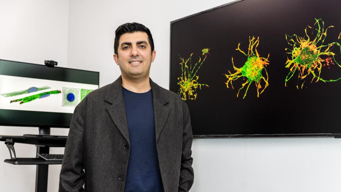
In a paper published this spring in Nature Materials, Alisafaei and a team of researchers at NJIT and Washington University in St. Louis described their discovery of a biomechanism that may be primarily responsible for the maladaptive response.
Fibroblasts, which are surrounded by a collagen structure known as the extracellular matrix, change little in shape or composition while performing regular tissue maintenance. When activated by an injury, they convert into specialized cells called myofibroblasts that generate high forces to pull the wound edges together. In doing so, they tug on the surrounding matrix and, in turn, feel resistance from it. This resistance creates mechanical tension that helps the cells sense the wound environment and adjust their activity accordingly.
The team learned that it is not just the amount of tension that matters in wound healing, but the direction in which it’s applied. To better understand these dynamics, they examined interactions between human dermal fibroblasts and their 3D fibrous matrix to quantify and model the two-way feedback loop between the two that determines these forces. Their results revealed that the directionality of mechanical signals from the extracellular matrix at early stages of an injury activates fibroblasts. They, in turn, remodel the matrix in a way that increases the tension in one direction more than others. It is this force, called anisotropy, that can malfunction if improperly regulated.
“By understanding how these mechanical signals drive cell behavior, we hope to develop better therapeutic strategies that enhance wound healing while preventing excessive scarring,” noted Alisafaei, an assistant professor of mechanical and industrial engineering.
One potential approach is to use adhesive strips to apply controlled tension to the skin. Fibroblasts naturally experience directional tension due to the alignment of collagen fibers along specific patterns known as Langer’s lines. Stretching the skin along these lines amplifies fibroblast activation, while applying tension in a different direction can suppress their activity. They are currently fine-tuning the amount of tension they would need in different directions to balance the forces and regulate the activation level of fibroblasts. Their next steps include developing wearable devices and biomaterials that can apply directional forces to wounds in a controlled way.
The team also considers the effects of aging and chronic disorders such as diabetes and obesity on fibroblast behavior. In these conditions, the extracellular matrix often becomes disorganized and mechanically compromised, making it more susceptible to anisotropic forces.
“When the matrix is less structured, fibroblasts can more easily realign its fibers,” Alisafaei explained. “This heightened sensitivity may help explain why patients with these conditions are more prone to excessive scarring or fibrosis. By better understanding how disease alters the mechanical landscape, we hope to design interventions that restore balance and promote healthy tissue repair.”