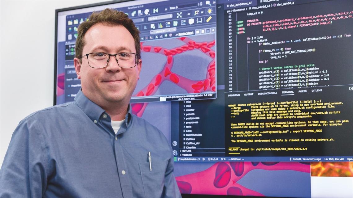
Threaded throughout the body are networks of capillaries so miniscule that red blood cells must contort to squeeze through them one at a time. It is this world that Peter Balogh brings to dynamic, 3D life on computer screens.
“The most important biological processes associated with blood flow occur in the tiniest vessels. This is where oxygen is exchanged between red blood cells and tissues, where new arteries sprout and where cancer cells enter tissues and metastasize,” says Balogh, a mechanical engineer at New Jersey Institute of Technology who specializes in computational fluid dynamics.
He has written new code that runs on supercomputers which allows him to model biological cells as they move through complex geometries, simulating the physics of bodily operations that experimental biologists can’t see. The capillaries he studies range in size from 5 to 50 microns in diameter. Blood cells, by comparison, are about 8 microns wide.
One of the forces Balogh studies is the shear stress that blood flow exerts on the walls of these vessels, which directly influences the growth of new vessels. In tissues that lack oxygen, the vessel wall senses the force and coordinates with chemical growth factors, causing new vessels to form. In the case of an artery blockage, the body may revascularize to improve blood flow around the blocked region. Diseases such as diabetes, however, inhibit this new growth, impairing the delivery of oxygen and nutrients that feed the retina, for example, which can lead to blindness.
“Our high-resolution images, coupled with the simulations, give a better idea about what promotes their formation and what prevents it. The accuracy in modeling these forces goes up in 3D,” he notes. “We try to determine why there is growth in one region and not in another.”
Red blood cells are remarkable in that they’ve evolved to not break. When they become stiff, as in sickle cell anemia, they are no longer able to flex in order to flow through capillaries and the narrow openings in the spleen. Backed up, the spleen becomes swollen and painful.
We can model the flow of any blood cells, including those trying to squeeze through and failing.
Balogh is currently collaborating with biologists at the University of Florida who send him high-resolution images of capillary networks in laboratory rats with particular disorders. He then reconstructs them digitally and models their blood cell flow to understand at a root level what the forces on the wall look like.
With patient-specific images of retinal capillaries in people with diabetes, he would be able to identify key differences in how blood flows through them compared to healthy networks. A further goal is to simulate the transport of drug particles with blood cells to see how they’re distributed through the retina, or in the case of sickle cell anemia, whether they can get through at all.
“These networks vary from one patient to another and so simulations would allow us to model the effects of a drug before we try it,” he notes. This could be a key tool in devising therapies for solid tumors, which he describes as “nasty and chaotic,” and thus even more patient-specific.